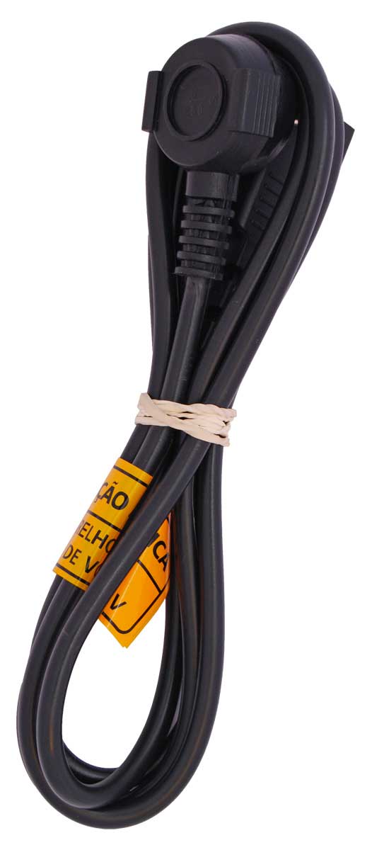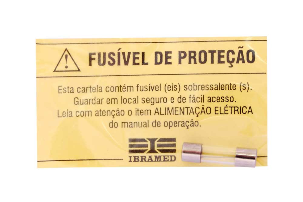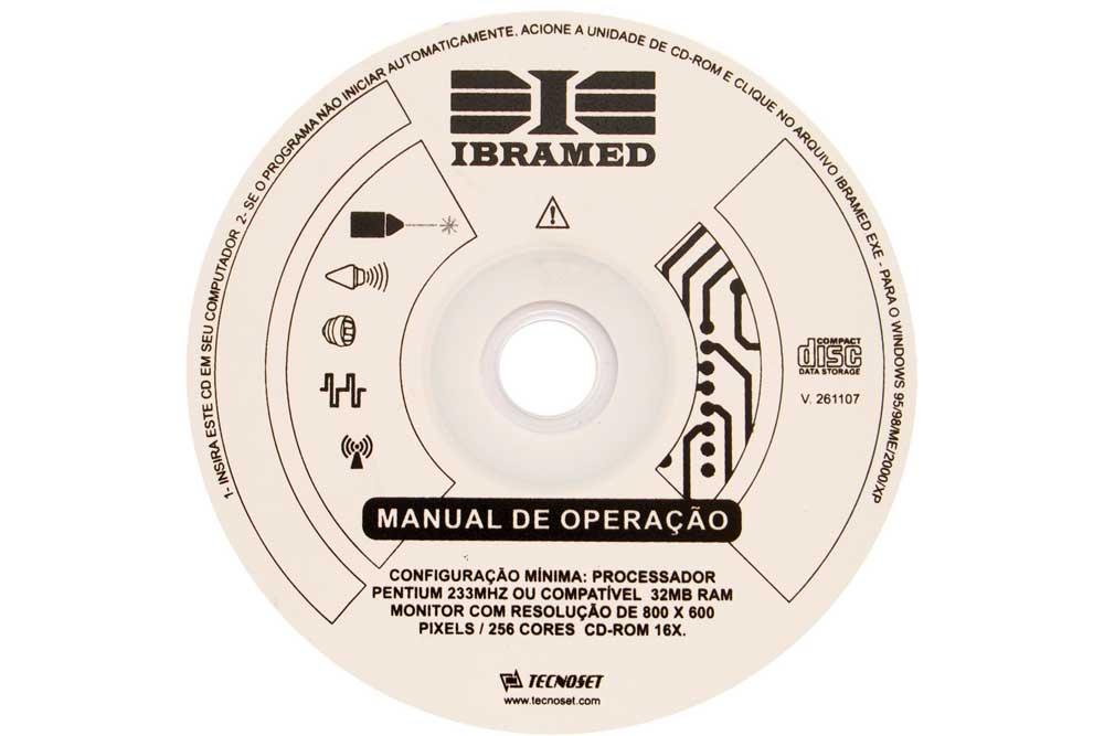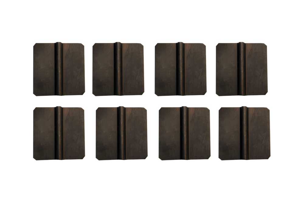
Neurodyn
- Motor stimulus
- Sensorial stimulus
- Rehabilitation
The equipment
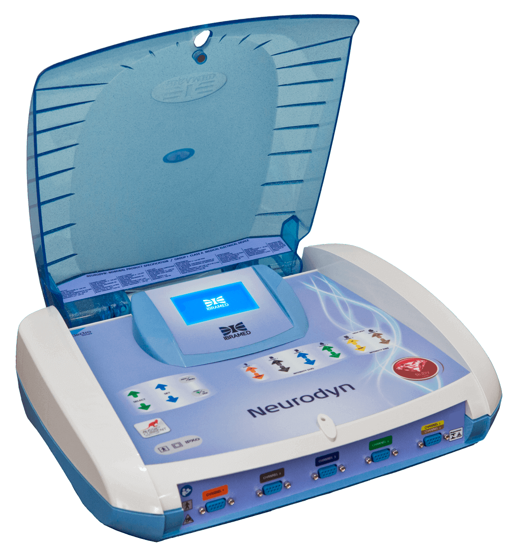
Neurodyn transcutaneous neuromuscular stimulator is a four-channel stimulator with independent controls for electrical current therapy using: Aussie Current (Medium Frequency Current modulated in Burst), Russian Current (Medium Frequency Current modulated in Burst), FES Current (Functional Electrical Stimulation),TENS Current (Transcutaneous Electrical Nerve Stimulation), Interferential Current (IFC-T) Tetrapolar (Medium Frequency Current modulated in Amplitude), Premodulated Interferential Current (IFC-B) Bipolar (Medium Frequency Current modulated in Amplitude), and with two independent control channels for the treatments using: Microcurrent (Microcurrent Electrical Neuromuscular Stimulation), Direcct Current/Polarized Current and High Voltage Pulsed Current (High Volt Pulsed Current).
The equipment is for use only under the prescription and supervision of a licensed professional.
Technical features
Electric Descripition
Input: 100 – 240 V ~ 50/60 Hz
Input power: 85 VA
Electric class: CLASS II

Aussie Current
Output mode: Electrodes
Intensity (CC): 0-120 mA* ±20%
Frequency of carrier (Carrier): 1 or 4 kHz ±10%
Duration of Burst (Burst ms): 2 or 4 ms ±10%
Current Mode
Continuous (Cont) 1, 2, 3 or 4 channels
Synchronous (Sync) 1, 2, 3 or 4 channels
Reciprocal (Rec) 1 and 3; 2 and 4 channels
Sequential (Seq) 1, 2, 3 or 4 channels
Frequency of Burst (Burst Hz): 1-120 Hz ±10%
Ramp
Duration of up ramp (Rise): 1-20 s ±10%
Duration of muscle contraction (On):1-60 s ±10%
Duration of down ramp (Decay): 1-20 s ±10%
Duration of muscle relaxation (Off): 1-60 s ±10%
Treatment duration (Timer): 1-60 min ±10%
Intensity Control (UP or DOWN): Individual channels of intensity 1, 2, 3 and 4
Russian Current
Output mode: Electrodes
Intensity (CC): 0-120 mA* ±20%
Current Mode
Continuous (Cont) 1, 2, 3 or 4 channels
Synchronous (Sync) 1, 2, 3 or 4 channels
Reciprocal (Rec) 1 and 3; 2 and 4 channels
Sequential (Seq) 1, 2, 3 or 4 channels
Duty cycle (Cycle): 10%, 20%, 30%, 40% and 50% ±10%
Burst Frequency Burst(Hz): 1-100 Hz ±10%
Ramp
Duration of up ramp (Rise): 1-20 s ±10%
Duration of muscle contraction (On):1-60 s ±10%
Duration of down ramp (Decay): 1-20 s ±10%
Duration of muscle relaxation (Off): 1-60 s ±10%
Treatment duration (Timer): 1-60 min ±10%
Intensity Control (UP or DOWN): Individual channels of intensity 1, 2, 3 and 4
FES Current
Output mode: Electrodes
Intensity (CC): 0-120 mA* ±20%
Current Mode
Synchronous (Sync) 1, 2, 3 or 4 channels
Reciprocal (Rec) 1 and 3; 2 and 4 channels
Frequency (Freq Hz): 0.5 – 250 Hz ±10%
Duration of pulse phase (Phase μs): 50-500 μs ±10%
Ramp
Duration of up ramp (Rise): 1-20 s ±10%
Duration of muscle contraction (On):1-60 s ±10%
Duration of down ramp (Decay): 1-20 s ±10%
Duration of muscle relaxation (Off): 1-60 s ±10%
Treatment duration (Timer): 1-60 min ±10%
Intensity Control (UP or DOWN): Individual intensity channels 1, 2, 3 and 4
TENS Current
Output mode: Electrodes
Intensity (CC): 0-120 mA* ±20%
Current Mode
Conventional or Continuous (Conv): Duration of pulse phase: adjustable from 50 -500 μs ±10%; Frequency: adjustable from 0.5 – 250 Hz ±10%.
Modulation in Burst (Burst Hz): Frequency fixed at 250 Hz ±10%; Frequency of Burst from 2 Hz ±10%; Duration of pulse phase: adjustable from 50 – 500 μs ±10%.
Automatic variation of intensity and frequency (VIF): 2 – 247 Hz ±10%; Duration of pulse phase: 50 – 500 μs ±10%; Frequency: 2 – 247 Hz ±10%
Duration of pulse phase (Phase μs): 50-500 μs ±10%
Frequency (Freq): 0.5 – 250 Hz ±10%
Treatment time (Timer): 1 – 60 min ±10%
Intensity Control (UP or DOWN): Individual Intensity Channels 1, 2, 3 and 4.
Interferential Current (IFC-T) Tetrapolar
Output mode: Electrodes
Amplitude (CC*): 0-120 mA* ±20%
Frequency of carrier (Carrier): 1, 2, 4, 8 and 10 kHz ±10%
Scanning mode (Scan):
Static
Automatic (Auto 10%, Auto 50%, Auto 100%)
Beat Frequency (AMF): 1-200 Hz ±10%
Sweep Frequency (Sweep Hz): 1-200 Hz ±10%
Sweep mode (Sweep s):
Off
1/1 s
1/5/1 s
6/6 s
Treatment duration (Timer): 1-60 min ±10%
Intensity control (UP or DOWN): Available in channels: 1 and 2 /3 and 4.
Current Mode
Synchronous (Sync) 1, 2, 3 or 4 channels
Reciprocal (Rec) 1 and 3; 2 and 4 channels
Frequency (Freq Hz): 0.5 – 250 Hz ±10%
Duration of pulse phase (Phase μs): 50-500 μs ±10%
Ramp
Duration of up ramp (Rise): 1-20 s ±10%
Duration of muscle contraction (On):1-60 s ±10%
Duration of down ramp (Decay): 1-20 s ±10%
Duration of muscle relaxation (Off): 1-60 s ±10%
Treatment duration (Timer): 1-60 min ±10%
Intensity Control (UP or DOWN): Individual intensity channels 1, 2, 3 and 4
Premodulated Interferential Current (IFC-B) Bipolar
Output mode: Electrodes
Intensity (CC): 0-120 mA* ±20%
Frequency of carrier (Carrier): 1, 2, 4, 8 and 10 kHz ±10%
Current Mode
Continuous (Cont) 1, 2, 3 or 4 channels
Synchronous (Sync) 1, 2, 3 or 4 channels
Sequential (Seq) 1, 2, 3 or 4 channels
Beat Frequency (AMF): 1-200 Hz ±10%
Sweep mode (Sweep s)
Off
1/1 s
1/5/1 s
6/6 s
Sweep Frequency (Sweep Hz): 1-200 Hz ±10%
Ramp
Duration of up ramp (Rise): 1-20 s ±10%
Duration of muscle contraction (On):1-60 s ±10%
Duration of down ramp (Decay): 1-20 s ±10%
Duration of muscle relaxation (Off): 1-60 s ±10%
Treatment duration (Timer): 1-60 min ±10%
Intensity control (UP or DOWN): Available in channels: 1, 2, 3 and 4.
Microcurrent(MENS)
Output mode: Electrodes
Output intensity (CC): 0 – 990 μA* ±20%
Polarity (Pole):
Positive (P+)
Negative (P-)
Alternate (Alt) (0.33 Hz ±10%)
Frequency (Freq): 0.1-500 Hz ±10%
Treatment duration (Timer): 1-60 min ±10%
Intensity control (UP or DOWN): Individual intensity channels: 1 and 2.
Frequency of current: 15 kHz ±10%
Current Mode
Synchronous (Sync) 1, 2, 3 or 4 channels
Reciprocal (Rec) 1 and 3; 2 and 4 channels
Frequency (Freq Hz): 0.5 – 250 Hz ±10%
Duration of pulse phase (Phase μs): 50-500 μs ±10%
Ramp
Duration of up ramp (Rise): 1-20 s ±10%
Duration of muscle contraction (On):1-60 s ±10%
Duration of down ramp (Decay): 1-20 s ±10%
Duration of muscle relaxation (Off): 1-60 s ±10%
Treatment duration (Timer): 1-60 min ±10%
Intensity Control (UP or DOWN): Individual intensity channels 1, 2, 3 and 4
Direct Current/Polarized Current (POL)
Output mode: Electrode
Output intensity (CC): 0 – 30 mA* ±20%
Frequency: 15 kHz ±10%
Polarity (Pole):
Positive (P+)
Negative (P-)
Treatment time (Timer): 1-60 min ±10%
Intensity control (UP or DOWN): Available in channels: 1 and 2.
High Voltage Pulsed Current (HVPC)
Output mode: Electrodes
Intensity: 0 – 400 Vp* ±20%
Frequency (Freq): 1 – 250 Hz ±10%
Polarity (Pole):
Positive (P+)
Negative (P-)
Current Mode
Continuous (Cont) 5 or 6 channels
Synchronous (Sync) 5 or 6 channels
Ramp
Duration of up ramp (Rise): 1-20 s ±10%
Duration of muscle contraction (On):1-60 s ±10%
Duration of down ramp (Decay): 1-20 s ±10%
Duration of muscle relaxation (Off): 1-60 s ±10%
Treatment Duration (Timer): 1-60 min ±10%
Direct Current/Polarized Current (POL)
Output mode: Electrode
Output intensity (CC): 0 – 30 mA* ±20%
Frequency: 15 kHz ±10%
Polarity (Pole):
Positive (P+)
Negative (P-)
Treatment time (Timer): 1-60 min ±10%
Intensity control (UP or DOWN): Available in channels: 1 and 2.
Aussie Current
Aussie current is an alternate current that realizes transcutaneous neuromuscular stimulation using medium frequency (4000Hz or 4 Khz) modulated in bursts, with short duration pulses; offering then a minor sensorial discomfort during therapy.
The therapy set to 1Khz with 2 ms of duration is used for muscular torque gain treatment, prevention or atrophy delay due to disuse, motor re-education, maintenance or increase of amplitude from articular movement. The bursts set to 4Khz with 4ms of duration used in the treatments for symptomatic relief of: intractable chronic pain, post traumatic acute pain, postoperative acute pain and increasing local blood flow.
Russian Current
The Russian current is defined as an alternate current of medium frequency (2.5 kHz), which provides a greater depth range, due to its lower resistance, thus contributing to a greater recruitment of motor units without significant discomfort to the patient. Their bursts have rectangular or sinusoidal shapes. It is an excitomotor current that uses surface electrodes for its application. The mechanism of action is the depolarization of the lower motor neuron, generating muscular contraction in the muscles that present their full motor innervation. It is used in therapies whose objectives are: prevention or delay of atrophy due to disuse, motor re-education, increase of muscular torque, maintenance or increase in range of motion, relaxation of muscle spasms, increasing local blood flow
FES Current
The Functional Electrical Stimulation (FES / EMS) is a low frequency electrical current (approximately 1 to 100Hz), which has the following characteristics: alternate, depolarized, with symmetrical pulses.
This electrical stimulation generates an evoked potential of action that will act at the motor level, in a bidirectional way, leading to a depolarization of the motor neuron. This potential of action passes through the neuromuscular junction, reaching the motor neuron; as a result, the plastic membrane generates an action that will spread through the T tubules, thereby causing the release of calcium from the endoplasmic reticulum. This will then stimulate muscular contraction, by means of synchronous stimulation of the motor units. This synchronism leads to an efficient muscular contraction.
FES / SEM is used in rehabilitation for the purpose of preventing or delaying atrophy due to disuse, motor re-education, increasing muscular torque, maintenance or increase of joint movement amplitude, relaxing muscle spasms and increasing local blood flow.
TENS Current
The Transcutaneous Electrical Stimulation (TENS) is a low-frequency current, very common in rehabilitation for the purpose of analgesia.
A physiology of the action of TENS is explained by “floodgate theory”, where the transcutaneous electrical stimulus, activates the larger caliber mechanoreceptor fibers that send signals in the posterior horn of the spinal cord before smaller caliber fibers. Occurs then a depolarization of the gelatinous substance and a modulation of the stimulus in the spinal cord, which causes inhibition of the nocioreceptive stimulus. With the faster arrival of the proprioceptive stimulus in the posterior horn of the spinal cord, will occur activation of interneurons besides the release of opiate substances, such as: endorphins, dynorphins, enkephalins, noradrenaline and serotonin; these substances, acting as inhibitory neurotransmitters.
This way, TENS therapy is used in the symptomatic treatment of intractable chronic pain, relief of postoperative acute pain and increasing local blood flow
Interferential Tetrapolar and Bipolar
We have two Interferential currents, both of them are medium frequency sinusoidal that alternate between then, being modulated by amplitude. Its application can be carried out in bipolar (2 poles) or tetrapolar (4 poles, where at the intersection point occurs an interference); therefore, deeper tissues are reached without causing major discomfort.
It is a current widely used for the treatment of acute and chronic pains in different pathologies, such as: low back pain, osteoarthritis of the knee and others, this analgesic effect being explained by the “floodgate theory”. In addition, the literature points to the use of Interferential Current for urinary incontinence, intestinal constipation and increasing local blood flow.
Microcurrent
Microcurrent therapy has an electric current provided by one millionth of an ampere, literally, because this is believed to be the body’s own natural current strength. This therefore restores the body’s own natural current (Mercola & Kirsch, 1995).
Microcurrent therapy comes with two self-adherent active electrode patches linked by a cable. The efficacy of Microcurrent therapy in the management of musculoskeletal pain and enhancement of wound healing has been reported.
It works on a cellular level to stimulate the healing process. It is based upon the theory that the body’s electrical balance is disrupted when one is injured, so that the natural electrical current of the body changes its course. Microcurrent stimulation restores this balance (Kirsch, 2001).
In fact, Microcurrent therapy can relieve pain, stimulate wound healing, stimulate the regeneration of injured tissue, provide relief to myofascial trigger points, increase.
Using Microcurrent therapy, ATP production increases by 500%. ATP is the primary molecule that our bodies use to produce energy and you can fiund in every cell of the body. In fact, it has been found that ATP production increased fivefold after microcurrent therapy was administered. As stated previously, protein synthesis also increased, and so did amino acid transport (Mercola & Kirsch, 1995).
In addition, Microcurrent therapy can be used at specific frequencies for a variety of tissues and conditions. This can often soften tissue and decrease pain, which provides long-lasting pain relief that may even be permanent. This has some promising benefits that may be applicable to current chronic pain conditions as well (Neumann, 1993).
Polarized Current
Iontophoresis or ionization, also called ion transfer, is the penetration of substances via transdermal (skin) in a conservative manner, using a polarized current that drives the ionized substance by unidirectional electrical flow. The success of ion transfer is on the basic physical principle, which confirms that similar poles repel and opposite poles attract each other.
The philosophy of iontophoresis is similar to sonophoresis, although, in this case, the active principle (pharmacology) incorporated to the gel (donating solution) must have polarity defined, it means, must be ionized. The drug is propelled by electro- repulsion mechanism, positive or negative equivalent drugs are liberated, as long as they are placed under the electrode which presents the same electric charge.
It is important to highlight that the pharmacology applied in this way have superficial and in loco action and must have essential characteristics such as hydro solubility, being positively or negatively charged and be of a relatively small molecular size (Oliveira et al., 2005; Cameron, 2009).
Like with sonophoresis, iontophoresis has potential routes of skin permeation, the sweet glands being the main way, while the porous follicle, the sebaceous glands and the corneous extract contribute relatively to the ionic penetration, since they have elevated relative electrical impedance (Oliveira et al., 2005).
A polarized electric current of constant amplitude (0.1 a 1 mA/cm2) is used to increase in a controlled way the transdermal transferring of the drug and in any type of equipment the electrode which will transfer the drug is called active and the other electrode which completes the electric circuit is called dispersive (Oliveira et al., 2005; Cameron, 2009).
High Volt Current
The high volt pulsed current (HVPC) is a current with twin pulses of high amplitude (high voltage) and short duration. The waveform is monophasic (the current flows in a single direction). The high volt causes a decrease in skin resistance making the current comfortable and tolerable. The system generally have a pulse duration that can vary from 5 to 200 µs (microseconds), a high peak amplitude (100 V to> 500 V) and frequencies of 1–250 Hz, allowing a relatively pleasant stimulation, capable of reaching the sensory, motor nerve fibers and also those responsible for conducting nociceptive impulses. Although HVPC is a single-phase current, the risks inherent in its use are minimized by the fact that the average amplitude of the direct current is very low, reaching values that can vary from 1.2 to 1.5 mA and a load that varies between 12 to 14 µC. Both, the average amplitude of the direct current and the load values, maintain a relationship with the large pulse intervals, which represent 99% of each second. HVPC produces a strong electromagnetic force that produces a “galvanotaxis effect” that induces the migration of polarized cells to the cathode or anode. The migration of neutrophils, macrophages, lymphocytes and platelets is believed to stimulate the “inflammatory phase” of the tissue healing response. Subsequent migration of fibroblasts to the wound site increases the synthesis of new tissue during the “proliferative phase” of the healing response. The migration of epidermal cells and keratinocytes is beneficial during the “remodeling and final maturation phase” of the healing response
Indications
- Relaxation of muscular spasms
- Prevention or retardation of disuse atrophy in post-injury type conditions.
- Increase local blood circulation.
Muscle re-education.
- Maintenance or increasing range of motion.
FES CURRENT (FES)
Stimulation of leg and ankle muscles of partially paralyzed patients to provide flexion of the foot, thus improving the patient’s gait.
TENS CURRENT (TENS)
Symptomatic relief of chronic intractable pain.
- Post-traumatic acute pain.
- Post-surgical acute pain.
INTERFERENTIAL CURRENT (IFC-T) – 4 POLES
Symptomatic relief of chronic intractable pain.
- Post-traumatic acute pain.
- Post-surgical acute pain.
PREMODULATED INTERFERENTIAL CURRENT (IFC-B) – 2 POLES
- Relaxation of muscular spasms.
Prevention or retardation of disuse atrophy in post-injury type conditions.
- Increase local blood flow
- Muscular re-education.
Maintenance or increasing range of motion.
- Symptomatic relief of chronic intractable pain.
- Post-traumatic acute pain.
- Post-surgical acute pain.
MICROCURRENT (MENS)
- Symptomatic relief of chronic intractable pain.
Post-traumatic acute pain.
- Post-surgical acute pain.
DIRECT CURRENT/POLARIZED CURRENT (POL)
- Relaxation of muscular spasm.
HIGH VOLT PULSED CURRENT (HVPC)
- Relaxation of muscular spasm.
- Prevention or retardation of disuse atrophy in post-injury type conditions.
- Increase local blood flow.
- Muscular re-education.
Maintenance or increasing range of motion.
Powered muscular stimulators should be used only under medical supervision as an adjunctive therapy for the treatment of medical diseases and conditions.
Contraindications
Neuromuscular stimulators must not be used in patients with cardiac pacemakers on demand or any other implanted electronic device.
This device must not be used for relief of local symptomatic pain of unknown etiology, unless a pain syndrome is diagnosed.
The stimulation must not be applied over the nerves of the carotid sheath, particularly in patients with altered sensitivity to the reflexes of the carotid sheath.
Stimulation must not be applied over the cardiac area, for it may cause cardiac arrhythmia.
Stimulation must not be applied over the skull.
Stimulation must not be applied over or near cancerous lesions.
Stimulation must not be applied over infected or inflamed areas, or skin eruptions such as phlebitis, thrombophlebitis.
Stimulation must not be applied over large caliber varicose veins, due to the risk of displacement of the thrombus.
Stimulation must not be applied in patients with deep venous thrombosis (DVT).
Stimulation must not be applied in the presence of electronic controlled equipment, (for example, cardiac monitors, ECG alarms), which may not function correctly when the electrical stimulation device is in use.
Warnings
- The safety use of neuromuscular stimulators during pregnancy has not been established.
- Care must be taken with patients suspected of cardiac conditions or conditions previously diagnosed.
- Stimulation must be applied cautiously over the anterior neck region. Muscle spasms of the larynx and pharynx may occur and the contractions may promote the closing of the upper airways, causing respiratory difficulties.
- Care must be taken in the treatment of patients suspected of or diagnosed with epilepsy.
- Care must be taken in the treatment with electrical stimulation in the presence of the following:
- When there is a tendency of hemorrhages after acute trauma or fracture;
- Following recent surgical procedures, when muscle contraction may disturb the healing process;
- Over the abdomen during pregnancy;
- Over areas of the skin where there is sensitivity deficit.
- The placement of electrodes and the configurations of stimulation must be in accordance with the prescriptions of the health professional.
- Neuromuscular stimulators must be kept away from the reach of children.
- Neuromuscular stimulators must be used only with those conductive wires and electrodes recommended by the manufacturers.
- Electrical stimulation with Polarized current must not be applied in areas of the skin which present lesions, because that region presents compromised dielectric impedance, and consequently the density of current will be greater. The high current may produce more lesions.
Adverse reaction
Patients may present skin irritation or hypersensitivity caused by electrical stimulation or electrically conductive medium. The irritation can usually be reduced with the use of an alternative conductor medium or with the placement of an alternative electrode.
- Patients may present burns under the stimulation electrodes if the application is not correctly carried out or if the electrodes are worn out.
Application techniques
The electrodes placement can be performed using the bipolar or monopolar technique. The adequate positioning and contact of the electrodes will assure comfort and efficacy of treatment.
Examine the skin for any wounds and clean the treatment area by rubbing the skin with medical grade alcohol.
Remove the self-adhesive electrodes from the protective backing and apply to the treated area as prescribed.
Make sure that the entire surface of the electrode is in contact with the skin of the patient, pressing it in place
Check the electrode contact regularly during treatment.
Examine the skin again and after the treatment.
Accessories
